|
|
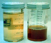
Subtopic 1: Physical Examination of Urine All routine urinalysis should begin with a physical examination of the urine sample. This examination includes assessment of volume, odor, and appearance (color and turbidity). Volume Urinary volume is dependent upon fluid intake; amount of solutes to be excreted, primarily sodium and urea; loss of body fluids by normal processes, such as perspiration and respiration, and abnormal processes, such as diarrhea; and cardiovascular and renal function. Although the volume of a random specimen is clinically insignificant, the volume of specimen received should be recorded for purposes of documentation and standardization. Urine volumes can be measured two ways: volumetrically and gravimetrically. That is, the volume is measured with a volumetric cylinder, or the volume is estimated by weighing the urine sample in a tared container and assuming that 1g = 1mL of urine. Odor Non-pathological, fresh urine has an inoffensive odor. One usually determines the odor of the urine sample by placing ones nose near the orifice of the sample container , moving the air from the container to your nose by gently wafting with your hand, and gently breathing the fumes. Appearance (color and turbidity) Color—The color of urine is related, to a large degree, by its degree of concentration. The color of non-pathological urine varies widely from colorless to deep yellow; the more concentrated the urine, the deeper the color. The color of urine is usually described after visual inspection with common color terms. Very often color charts will be available to report the colors in a consistent fashion. A good clinical history can resolve possible causes of an unusual urine color.
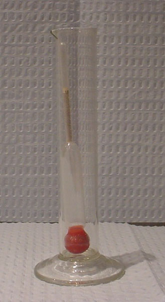
Turbidity—Normally freshly voided urine is clear. When urine is allowed to stand, amorphous crystals, usually urates, may precipitate and cause urine to be cloudy. The turbidity of urine should always be recorded and microscopically explained. Specific gravity—A hydrometer (urinometer) and a suitable container may be used to determine specific gravity.
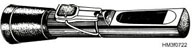
Many laboratories may also be equipped with refractometers that can relate density of a solution to specific gravity. Refractometers work on the principle that light passing from a transparent medium of one density to a medium of another density, will change its velocity and therefore the direction in which the beam of light is moving.
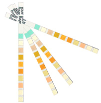
An indirect colorimetric method for estimating specific gravity is available on reagent strips ("urine dipsticks"). This method uses a pad that contains a complex, pre-treated electrolyte that undergoes a pH change based on the ionic concentration of the urine. This change results in a change of color of the pad. For the Multistix SG-10, specific gravity is measured using an apparent pKachange in the presence of an indicator (dyes) whose colors vary from deep blue-green at low ionic strength to green and yellow-green at higher ionic concentration. This estimate of specific gravity is rapid, simple, and requires no special equipment. The falling drop method is a direct method of measuring specific gravity that is usually used with automated instruments, such as the Clinitek Auto 2000 (Ames Division, Miles Laboratories, Inc., Elkhart , IN). The CliniTek2000 uses a specially designed column containing silicone oil. Specific gravity is calculated from the time it takes for a drop of urine to fall between two optical gates. Osmolality—Osmolality is usually measured by an osmometer, most frequently by a freezing point osmometer. Osmolalityis a measure of the number of particles per unit mass, whereas the specific gravity is a reflection of the density (mass per unit volume) of the suspended particles. Clinical Significance—Primary kidney function includes the ability to produce, in the appropriate circumstances, either a concentrated urine (osmolality>850 mOsm/kg) or a dilute urine (osmolality<100 mOsm/kg). A random urine whose osmolality>600 mOsm/kg is presumptive evidence of an ability to concentrate urine. The urine osmolality thus is part of the mechanism of maintaining water balance. In the presence of excess free water, the kidneys will produce a dilute urine, while in periods of water lack, a concentrated urine is produced. Loss of concentrating ability is often one of the earliest signs of kidney disease, clinically evidenced as nocturia (needing to void at night) and polyuria (increased volume of [usually dilute] urine). |