Activity
2 - Instructor Teaching Strategies
IDENTIFICATION OF PHI X 174 RF DNA BY RESTRICTION MAPPING
SUGGESTED TEACHING STRATEGIES
• Level of
difficulty: intermediate
• Students will need printed copies of the procedures.
• Students can work on the NEBcutter internet activity while their gels
are running, or it may be given as homework to print out before the lab. The
NEBcutter activity can also be given as a stand-alone activity without the wet
lab.
• Optional: DNA fragment sizes may be determined exactly by running 2
mg l/HindIII DNA size
markers (in a 10 ml volume) plus 1 ml
6x gel loading dye in a neighboring lane. It is important to heat the l/HindIII
DNA markers before loading them on the gel (70 ’ C for 5 minutes), otherwise
some of the DNA marker fragments will stick together, causing an unexpected
banding pattern
• This activity assumes a general knowledge of DNA structure.
• If students have trouble visualizing how a circular piece of DNA becomes
a series of linear fragments, have them model the digests using a long, skinny
piece of paper. Have them mark the coordinate 0 on one end, 5386 on the other
end, and the restriction sites in between. Tape the 0 end to the 5386 end, and
use scissors to produce the fragments.
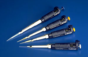
• Students will have to know how to use a micropipetter. Most manufacturers
have online instruction manuals. For an example, visit www.rainin.com, then
click on “literature”, and “Pipetman (single channel) manual”.
A separate student lab on micropipetter use may be found at www.swmed.edu/stars/resources/stock01/gaona.html
(Image from http://www.fotodyne.com/epdmicro.html, 2003)
• For streaming
video segments with gel electrophoresis tips, visit http://www.carolina.com/biotech
and click on “electrophoresis teacher tips”.
• Non-toxic DNA stains, such as Bio-Rad’s Bio-SafeTM DNA stain (www.explorer.bio-rad.com),
or Edvotek’s DNA Blu InstastainTM (www.edvotek.com)
can be used instead of toxic Etidium Bromide. This may increase staining time,
and eliminates the need for a UV transilluminator. You will want a white light
box instead. Bio-Rad also sells gel support film to dry gels for a permanent
record. That would eliminate the need for a camera system.
• For a wealth of information about restriction enzymes and related terminology,
go to http://rebase.neb.com/rebase/rebhelp.html.
TIME
COMMITMENT
• The entire lab can be completed in a single 3 hour lab period.
• If desired, the digested DNA samples may be kept at 4’C before
running them on the gel. In that case it is a good idea to heat them first at
70’C for 5 minutes to heat-inactivate the enzymes. They can also be stored
at 4’C by simply adding the gel loading buffer.
• If desired, gels may be pre-cast and kept in the gel box covered with
1x TBE buffer at room temperature. Do not remove the comb until just before
use.
• If desired, gels may be wrapped in saran wrap after running, and kept
at 4’C
overnight. Keep in mind this will cause the bands to appear less sharp
• Counting plaques takes 15-30 minutes
• Plates should be taken out of the incubator after the overnight incubation.
If desired, cover the rim with a strip of parafilm. Sealed plates can be stored
at 4’C for several weeks before counting.
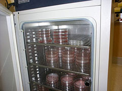
(Image from www.alz.uci.edu/Biochem1.html, 2003)
MATERIALS
Phi Use only autoclaved, distilled water (double-distilled preferred)!
• 1x TBE buffer (may be purchased as 10X TBE buffer from BioRad, catalog
# 161-0770EDU, or can be made from scratch by mixing 108 g Tris base and 7.44
g EDTA (disodium) in ~ 900 ml distilled water, then add 55 g boric acid and
bring the total volume up to 1 liter; adjust to pH8)
• 10x NEB 4 buffer (comes with New England Biolab restriction enzymes)
• Electrophoresis-grade agarose
• 6x gel loading dye (to make your own 6X solution, mix 250 ml
of a 1% solutions of Bromphenol blue, 250 ml of a
1% solution of xylene cyanol, 7 ml of 1 M Tris-HCl
(pH8), 443 ml distilled water, and 300 ml
glycerol. Alternatively, BIO Systems101 sells a 10X ready-made gel loading dye:
catalog # 2333-104).
• DraI restriction enzyme (e.g. New England Biolabs, catalog # R0129S,
2000 Units/100 ml)
• MfeI restriction enzyme (e.g. New England Biolabs, catalog # R0589S,
500 Units/50 ml)
• PhiX174 RFI DNA (e.g. New England Biolabs, catalog # N3022S, 30 mg/30
ml)
• Optional: l/HindIII DNA size marker e.g.
New England Biolabs, catalog # N3012S, 150 mg/300
ml)
• Stock Ethidium Bromide(kept in a dark bottle) - dilute to 0.5
mg/ml to stain. Ethidium Bromide solutions must be
treated with bleach before discarding with copious amounts of water in the sink,
or they can be disposed of with BIO101Systems’ EtBr Green BagTM kit (catalog
# 2350-200, http://www.qbiogene.com).
• sterile micropipetter tips
• 1.5 ml or 2.0 ml sterile microcentrifuge tubes
• laboratory labeling tape for taping up the gel tray
EQUIPMENT
• micropipetter
• sharpie pens
• hot plate or microwave
• protective gloves for handling hot agarose
• horizontal minigel box
• power supply
• microcentrifuge
• ice bucket or cold block
• 37’C water bath
• 70’C water bath or heat block
• two plastic trays
• 1 large spatula
• UV illuminator and protective face shield (if EtBr is used)
• Latex gloves (if EtBr is used)
• Polaroid camera with attached hood and type 667 polaroid film
• Computer with internet access and printer
TROUBLESHOOTING TIPS
• Bands larger than 5.39 kb on the gel are probably due to undigested,
still circular DNA. Any incomplete (“partial”) digestion will cause
extra bands to appear. First try increasing the incubation time to 2 hours.
If that does not work, double the amount of enzyme, or purchase fresh enzyme.
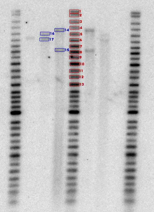
(Image from www.scanalytics.com/product/oneD/1dmac_gel.shtml, 2003)
•
If a long smear appears on the gel, the DNA was degraded by endonuclease enzymes.
Endonucleases come from hands and can end up in stock solutions.
• No bands may indicate an insufficient amount of DNA. Alternatively it
may indicate insufficient staining, or overstaining followed by insufficient
destaining.
• Beware tiny fragments may run off the gel into oblivion when run too
long.
• If bands are distorted, the gel may have been run too fast. Lower the
voltage, or cool the gel with a small fan during the run. Alternatively, there
may have been too much buffer covering the gel.
• Enzymes must be kept in a -20’C NON-frost-free freezer as
much as possible, or they will gradually lose their activity.